Haase Awarded $2.2M NIH MIRA ESI Grant to Study Cellular Dynamics
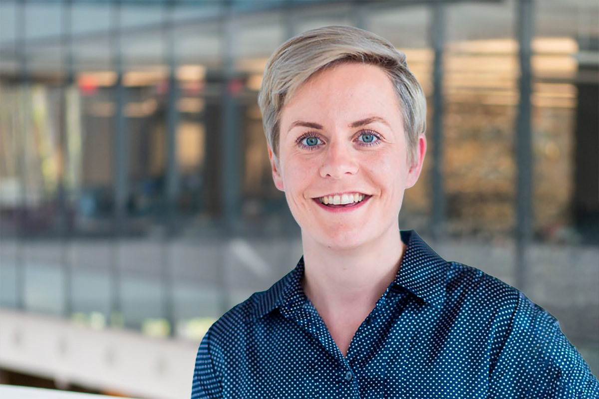
Christa Haase, assistant professor at Northeastern University jointly appointed in Bioengineering and Physics, has been awarded a five-year, $2.2 million R35 MIRA (Maximizing Investigators’ Research Award) from the National Institute of General Medical Sciences (NIGMS), part of the National Institutes of Health. The award, granted through NIGMS’s Early Stage Investigator (ESI) program, will support Haase’s research program, “From Motion to Mechanism: How Cellular Dynamics Shape Molecular Programs and Function In Vivo.”
The Haase Lab will build on its previously developed platform, Image-seq, which combines high-resolution live imaging with spatially resolved single-cell transcriptomics—allowing researchers to link a cell’s molecular identity to its dynamic behavior in live animals. The new funding will expand the technology in two directions: adding single-cell proteomics to capture protein-level signaling and applying it to axolotl limb regeneration using a custom three-photon microscope capable of deep tissue imaging. The axolotl work aims to produce the first-ever 5D (space, time, and cell cycle) single-cell map of early regeneration.
Together, the project’s new tools—including “Image-seq v2” and “Image-seq 3P”—are expected to shed light on how cell migration, proliferation, and signaling shape development, immune responses, disease, and regeneration across species, with potential implications for future therapeutic strategies.
The project runs from September 2026 through May 2031.
Abstract Source: NIH
Project Summary Cellular dynamics such as migration, proliferation, and signaling transients are central to tissue development, regeneration, immune function, and multicellular organization. These behaviors are also tightly linked to pathological states, including uncontrolled growth and metastatic spread, yet the molecular mechanisms by which distinct dynamic states influence intercellular communication remain poorly understood. Existing single- cell and spatial multi-omics technologies provide detailed molecular information but are limited to static snapshots, while intravital microscopy can capture dynamic behaviors but lacks molecular resolution. To address this gap, we previously developed Image-seq, a platform that integrates high-resolution in vivo optical imaging with image-guided, spatially resolved single-cell transcriptomics, thereby preserving both the spatial and temporal context of analyzed cells. Image-seq uniquely links molecular information to the dynamic history of cells captured by live imaging. Despite these advances, key limitations remain: (i) Image-seq is currently optimized for RNA sequencing, whereas much of intercellular communication is mediated at the proteomic level; (ii) Image- seq has been paired with confocal and two-photon microscopy, which offer limited penetration depth in scattering tissues; and (iii) applications have been confined to the mouse bone marrow, despite the potential for transformative insights across diverse tissues and vertebrate systems. To overcome these limitations, we propose to expand Image-seq in three major directions. First, we will integrate Image-seq with single-cell proteomics to directly capture protein-level signaling events, and define how distinct proliferative states shape intercellular communication within the bone marrow. Second, we will adapt Image-seq for use in the brain, where we will track immune cell infiltration longitudinally and correlate infiltration kinetics with rewiring of local signaling pathways. Third, we will extend Image-seq to axolotl regeneration, combining it with a custom-built three-photon and third-harmonic generation microscope that provides increased imaging depth, label-free structural contrast, and compatibility with large 3D tissue volumes. This will enable generation of the first 5D (x, y, z, t, cell cycle) single-cell map of early axolotl regeneration using continuous time-lapse intravital imaging. Together, these studies will deliver new technologies – including image-guided single-cell proteomics (Image- seq v2), three-photon Image-seq (Image-seq 3P), and artificial intelligence/machine learning pipelines for dynamic feature discovery – and provide fundamental insights into how distinct cellular dynamics shape transcription, translation, intercellular communication, and functional outcomes. In the long term, these advances are expected to yield broadly applicable frameworks for studying development, immune responses, disease, and regeneration across vertebrate classes, from mammals to amphibians.