Imaging the Brain With Transparent Array of Microelectrodes
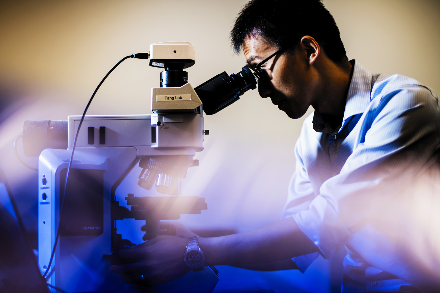
ECE Assistant Professor Hui Fang and a team of neuroscientists from Boston Children's Hospital have developed a transparent array of microelectrodes on nano-mesh to monitor the impulses sent by the brain.
Source: News @ Northeastern
Chain-link fences are common, and for good reason: They’re simple and flexible, without blocking light or visibility. As Hui Fang and a team of neuroscientists from Boston Children’s Hospital reveal in a new report published Wednesday in the journal Science Advances, their structure can also work wonders for the brain.
“I’m not a neuroscientist—and you’re probably not, either,” said Fang, an assistant professor of electrical and computer engineering at Northeastern. “But we still know that there are electrical impulses from neurons.”
Your neurons are firing as you read this, and researchers have the ability to monitor those impulses by implanting tiny electrodes directly onto the brain—the “gold standard” of mirroring fast brain activity, as Fang put it.
These electrodes range in size and flexibility, yielding to the contours of the brain in search of a signal. But with a subject as complex as the brain, even electrodes don’t tell the whole story.
“With only electrodes, you can’t tell sophisticated spatial information,” said Fang, listing a neuron’s shape, type, and connections as examples of data that fall through the cracks. “But that’s where optical methods can play a big role.”
While electrodes pick up impulses as they happen, optical tools acquire their own signals. In optical imaging, researchers shine low-level light into the brain, which can reveal detailed spatial information about the cells.
Since optical imaging is the missing link to revealing finer details, many researchers have begun to question the value of using electrodes as a standalone method. Bridging electrical activity and visuals, said Fang, is what will paint a full picture.
work in his lab.
Photo by Adam Glanzman/Northeastern University
However, a standard array of microelectrodes is opaque, making simultaneous imaging very difficult. Its metal layers and signal-boosting coating block out the light, which also makes it hard to use light to stimulate neurons.
The solution? Poke holes.
More specifically, Fang and his team opted to transform standard microelectrode materials into nano-mesh, a surface perforated by holes so small that they’re invisible even through a microscope.
“We’re using basically the same electrode materials as in conventional, non-transparent—and even rigid—electrode arrays,” said Fang. But by reconceiving the structure of the materials, his team found a way to make the electrode units not only soft and small, but see-through.
When lined up side by side, these tiny holes render the material transparent. The electrodes’ substance and stability come from the remaining materials, just like in a chain-link fence.
Not all materials were up to the challenge, though. In some cases, the coating covered the holes on the metal mesh. Fortunately, the polymer coating that the team ultimately chose withstood the modifications better than they could’ve imagined.
“Somehow, magically—we don’t fully understand the chemistry yet—it can maintain the same mesh structure,” said Fang.
The team soon tested their design in the lab, implanting the arrays on the brains of live mice. As the mice responded to visual stimuli, the researchers were able to produce high-resolution brain images, all while electrodes successfully traced the electrical activity back to individual neurons.
Fang’s electrodes—each only a few times as wide as a human hair—sit in sets of 32, but this coverage still pales in comparison to the scope of brain activity. With the eventual goal of use on humans, the team must first scale up each array’s capacity from a few dozen electrodes to thousands.
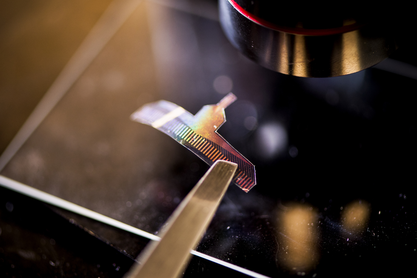
array in Fang’s lab.
Photo by Adam Glanzman/Northeastern University
Human trials could start in as few as three to five years, but the future stages of the technology are still uncertain. “I don’t have a crystal ball,” joked Fang, “so I don’t have a good prediction.”
It will likely take even longer for these arrays to be ready for use in children, whose brains are constantly developing. Eventually, though, Fang predicts that these devices will help researchers and other professionals deepen their understanding of conditions such as epilepsy and concussions in brains of any age.
In fact, the group has already begun working with Tufts University to identify new biological markers of traumatic brain injury. For now, though, their focus is fine-tuning the technology, combining their knowledge of neuroscience and engineering as they progress toward their goal.
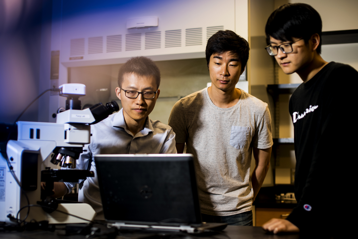
Fang described how the team of researchers at Northeastern and Boston Children’s Hospital, including two Northeastern graduate students, regularly discuss the technical difficulties of the project and the details of animal surgery. “Although they’re neuroscientists, they’re also very interested in technology development,” said Fang. “This collaboration is one of the best I have had in my career.”
Not much separates Northeastern and Boston Children’s Hospital anyway—only a 20-minute walk and a few chain-link fences. Plus, with transparent microelectrodes, the future is looking bright.