3D Printing Could Help in Finding Brain Cancer Treatments
BioE Associate Professor Guohao Dai, who specializes in 3D printing live tissue, created a three-dimensional model to study the development of brain tumors.
A 3D-printed brain could make it easier to find cancer treatments
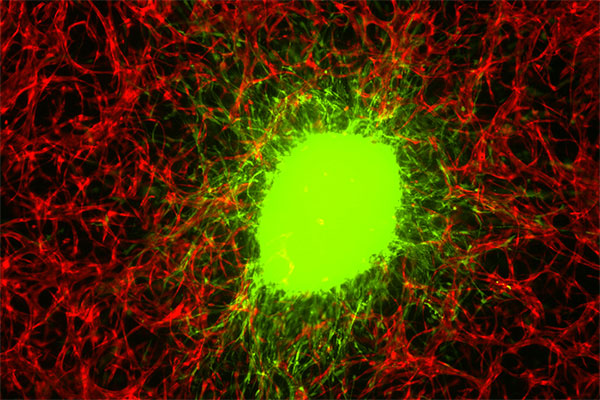
Photo: Glioblastoma stem cells aggressively invade a model made of human brain cells and biomaterials. Photo courtesy of Guohao Dai
Glioblastoma is the deadliest form of brain tumor—less than 10 percent of people who are diagnosed with it will survive more than five years.
A group of researchers has devised a new way to study this rapidly spreading cancer, using a three-dimensional structure made of an agglomeration of human brain cells and biomaterials. Their work, published in Science Advances, could help medical professionals better understand how the tumor grows and to speed up the potential discovery of new drugs to fight it.
“This is a very difficult brain tumor to treat,” says Guohao Dai, an associate professor of bioengineering at Northeastern and corresponding author on the study. “And it’s also difficult to do research on the brain tumor, because you cannot really see what’s happening.”

Guohao Dai is an associate professor of bioengineering in the College of Engineering at Northeastern. Matthew Modoono/Northeastern University
Inside a living brain, researchers can’t directly observe how tumor cells grow and respond to treatment. Studies are typically done in mice or rats, and the animals must be dissected to understand the tumor’s development. Animal studies are expensive and time-consuming, Dai says, and they don’t allow for day-to-day observations of the same tumor in living tissue.
To be able to study glioblastoma more directly, Dai, whose lab specializes in 3D printing live tissue, grew a three-dimensional model to act as brain tissue for tumor cells to infiltrate.
“We use human brain blood vessel cells, and connect them with all the neurons, pericytes, astrocytes, the major cell types in the human brain,” Dai says. A water-based substance known as a hydrogel serves as a matrix to hold these cells in place. “Then we use 3D printing to stack them in three-dimensional fashion.”
In the middle of the structure, which is only a few millimeters thick, the researchers place glioblastoma tumor stem cells. The cells are derived from brain tumor patients with the help of Jenny Zou, a neurosurgeon, and Roland Friedel, a neuroscientist, at Mount Sinai’s medical school.
“We can observe how the brain tumor cells aggressively invade, just like what we see in patients,” Dai says. “They invade everywhere.”
To get an accurate picture of what’s happening inside the 3D model without disrupting it, Xavier Intes, a biomedical engineer at Rensselaer Polytechnic Institute, used a laser to scan the sample and quickly create a three-dimensional snapshot of the cellular structure, an imaging technique developed in his lab.
This combination of techniques allowed them to evaluate the effectiveness of a commonly used chemotherapy drug, temozolomide.
“We treated the tumor with the same kind of drug you give to a patient when they undergo chemotherapy,” Dai says. “We monitored this chemotherapy over two months. And what we found was the chemotherapy was not able to kill the tumor.”
Initially, the tumor shrank in response to the drugs, but then it grew back swiftly and aggressively. The drug did not work in the long term, which lines up with the experience of patients with glioblastoma.
“This particular chemotherapy is not effective for the brain tumor,” Dai says. “We need to develop and screen other chemotherapy drugs.”
This model may be able to speed up that process. Temozolomide was able to kill glioblastoma cells in two-dimensional models, but when put into a three-dimensional one, the tumor rebounded. This method could be used to weed out unsuccessful drugs early, ensuring that only the most promising ones move to animal, and eventually human, trials.
“You have a tremendous amount of time and cost associated with animal research,” Dai says. “With our 3D glioblastoma model and imaging platform, you can see how the cells respond to radiation or chemotherapy very quickly.”
by Laura Castañón, News @ Northeastern